当教室から最近発表された原著論文をご紹介します。
Distribution and components of interstitial inflammation and fibrosis in IgG4-related kidney disease: analysis of autopsy specimens.
Hara S, Kawano M, Mizushima I, Harada K, Takata T, Saeki T, Ubara Y, Sato Y, Nagata M.
Hum Pathol. 2016 Sep;55:164-73. ![]()
2015 Impact Factor: 2.791
 IgG4関連疾患(IgG4RD: IgG4-related disease)は血清IgG4高値と組織へのIgG4陽性形質細胞の浸潤を特徴とする全身性の炎症性疾患です。腎臓は代表的な臓器病変のひとつでIgG4関連腎臓病(IgG4RKD: IgG4-related kidney disease)と呼ばれており、慢性化すると線維化を呈し、慢性腎不全へ至ります。これまでの研究によりIgG4RKDの特徴として被膜を越える炎症細胞浸潤、病変部と非病変部の境界明瞭な分布、storiform fibrosis (bird’s-eye pattern fibrosis)とよばれる特徴的線維化といった特異的病理学的所見が明らかになっていますが、これまでの研究は針生検という限られたサンプルでの解析であったことから、病変が実際にどのような広がりをもつのかは不明でした。また、storiform fibrosisが他の腎間質線維化とどのような違いがあるのかは明らかではありませんでした。そこで今回私たちは、IgG4RKDの剖検標本を用いて、IgG4RKDの病変の拡がりと線維化の成分を解析しました。
Abstract
IgG4-related kidney disease (IgG4-RKD) occasionally progresses to chronic renal failure and is pathologically characterized by IgG4-positive lymphoplasmacyte-rich tubulointerstitial nephritis with storiform fibrosis (bird’s-eye pattern fibrosis). Although radiology reveals a heterogeneous distribution of affected areas in this disease, their true distribution within the whole kidney is still unknown because of difficulty in estimating this from needle biopsy samples. Using 5 autopsy specimens, the present study histologically characterized the distribution and components of interstitial inflammation and fibrosis in IgG4-RKD. Interstitial lymphoplasmacytic infiltration or fibrosis was observed in a variety of anatomical locations such as intracapsular, subcapsular, cortical, perivascular, and perineural regions heterogeneously in a patchy distribution. They tended to be more markedly accumulated around medium- and small-sized vessels. Storiform fibrosis was limited to the cortex. Immunostaining revealed nonfibrillar collagens (collagen IV and VI) and fibronectin predominance in the cortical lesion, including storiform fibrosis. In contrast, fibril-forming collagens (collagen I and III), collagen VI, and fibronectin were the main components in the perivascular lesion. In addition, α-smooth muscle actin-positive myofibroblasts were prominently accumulated in the early lesion and decreased with progression, suggesting that myofibroblasts produce extracellular matrices forming a peculiar fibrosis. In conclusion, perivascular inflammation or fibrosis of medium- and small-sized vessels is a newly identified pathologic feature of IgG4-RKD. Because storiform fibrosis contains mainly nonfibrillar collagens, “interstitial fibrosclerosis” would be a suitable term to reflect this. The relation between the location and components of fibrosis determined in whole kidney samples provides new clues to the pathophysiology underlying IgG4-RKD.
要旨
多施設から集積したIgG4-RKDの剖検症例5例の腎臓標本を用いました。IgG4-RKDの病変を局在(被膜内・被膜下・皮質・血管周囲・神経周囲・髄質)と進展度(A-D)によって分類し、病変の分布と特徴的成分を免疫染色(Collagen type I, III, IV, VI, fibronectin, alpha-smooth muscle actin: αSMA)で解析しました。また対照群として非IgG4-RKDにおいても同様の免疫染色を行いました。
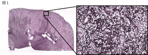
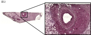
その結果、特徴的な分布として、多くのIgG4-RKDの病変は皮質または血管周囲に分布しており、特異的な線維化であるstoriform fibrosisは皮質に限局していることがわかりました(図1)。また比較的深い中型血管周囲の病変が初めて明らかになりました(図2)。特徴的な成分として、血管周囲病変ではcollagen Iを含めたcollagen III, IV, VI, fibronectinが認められた一方、storiform fibrosisを含む皮質病変ではcollagen Iは含まれておらずcollagen IV, VI, fibronectinが主体でした。一方、非IgG4-RKDではcollagen Iを含むcollagen III, IV, VI, fibronectinが主体でした。さらに筋線維芽細胞を示すα-SMA陽性細胞は早期で最も多く、病変が進展するほど有意に減少することがわかりました。
今後の展望
今回の研究でIgG4-RKDの新たな特徴的病理所見として中型血管周囲の病変が明らかとなり、IgG4-RDが血管外膜に沿って大型血管から中型血管にかけて分布する可能性が示唆されました。また、storiform fibrosisはcollagen Iを含まない基質主体の間質線維化であることから、非IgG4-RKDの間質線維化とは異なる病態と考えられました。これらの特徴的な病変の分布や成分がどのような機序で形成されるかを明らかにすることが今後の課題となります。それには動物モデルを用いた解析が望ましく、IgG4-RDに特異的な治療へ繋がる可能性があると考えています。


