当教室の大学院生でリウマチ膠原病内科専門医でもあります、覚知泰志先生の学位論文がModern Rheumatology誌に掲載されます。IgG4関連疾患に関しては、当教室が世界のオピニオンリーダーの一つに数えられるほど精力的な研究を進めています。今回は、IgG4関連疾患で多臓器に病変を発症した症例のIgG4陽性細胞の性格を分析しました。その結果、多臓器においても共通のクローンをしめす細胞が存在することがしめされています。
Kakuchi Y, Yamada K, Ito K, Hara S, Fujii H, Yamagishi M, Kawano M.
Analysis of IgG4-positive clones in affected organs of IgG4-related disease.
Mod Rheumatol. 2016 Mar 4:1-30. [Epub ahead of print]
http://www.ncbi.nlm.nih.gov/pubmed/26943245
Abstract
OBJECTIVE: We investigated class switch reaction (CSR) in affected organs and evaluated whether the same or genetically related clones exist in IgG4-RD.
METHODS: We studied three patients with IgG4-RD. Total cellular RNA was extracted from salivary glands and peripheral blood, and lung tissue. Activation-induced cytidine deaminase (AID) and immunoglobulin heavy chain third complementarity determining region (IgVH-CDR3) of IgM and IgG4 were detected by reverse transcription polymerase chain reaction. We analyzed the clonal relationship of infiltrating IgG4-positive cells, as compared with IgM. We determined the existence of common clones among organs and patients.
RESULT: AID was expressed in salivary glands of all patients and lung tissue in one. Closely related IgVH-CDR3 sequences in infiltrating IgG4-positive cells were detected in salivary glands and lung tissue. Identical IgVH-CDR3 sequence between IgM and IgG4 in salivary glands was detected in one patient, indicating CSR in salivary glands. Identical IgVH-CDR3 sequences of IgG4-positive cells were detected between salivary glands and peripheral blood in two patients. Four identical sequences of IgVH-CDR3 existed between patients. Interestingly, one of the four sequences was detected in all patients.
CONCLUSION: Our results demonstrate the existence of common antigen(s) shared by patients with IgG4-RD.
当教室では、臨床重視の方針から症例報告論文の執筆を積極的に奨励しています。今野哲雄助教は、本来心筋症など、遺伝性心血管疾患の臨床研究を進めています。今回は、肥大型心筋症によくみられる、心電図胸部誘導での巨大陰性T波類似の所見が、著明な低カリウム血症で認められた症例の記載です。巨大陰性T波の鑑別の一つとしての重要な所見と思われます。
Konno T, Hayashi K, Fujino N, Yamagishi M.
Hypokalemia and the Disappearance of Giant Negative T Waves.
Intern Med. 2016;55(5):545-6. doi: 10.2169/internalmedicine.55.5770. Epub 2016 Mar 1.
https://www.jstage.jst.go.jp/article/internalmedicine/55/5/55_55.5770/_article
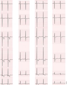
A 76-year-old woman with hypertrophic cardiomyopathy (HCM) was admitted to our hospital for hypokalemia (1.5mmol/L) due to diarrhea. An electrocardiogram taken 5 months before this admission, when the patient’s potassium level was 4.2 mmol/L, showed giant negative T waves (PictureA), whereas they were absent on this admission (PictureB). The potassium imbalance was corrected on the third hospital day. The amplitude of the negative T waves gradually increased, and deep negative T waves reappeared 2 months after the correction of the potassium level (Picture C). Three months after discharge, the patient was readmitted to our hospital with hypokalemia (2.4 mmol/L) due to recurrent diarrhea and again showed an absence of the giant negative T waves (Picture D). Drastic changes in the amplitude of the negative T waves in relation to the serum potassium levels in this case represent a role for serum potassium ions in cardiac repolarization because hypokalemia decreases potassium conductance and may cause flat T waves, even when classical electrocardiogram findings are observed in HCM The authors state that they have no Conflict of Interest


